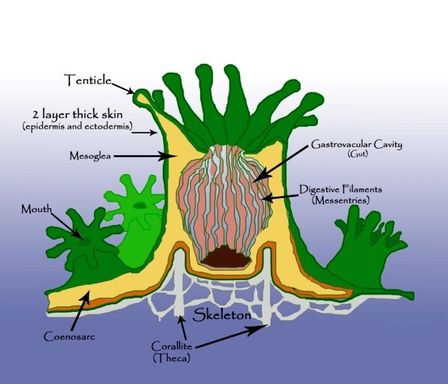
- Mouth - The opening on the oral disc that leads to the gastrovascular cavity and serves as both mouth and anus.
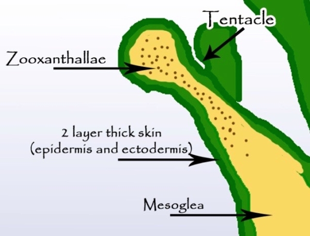
- Tentacles - Flexible, extendable structures around the oral disc, used for prey capture and defence, contain stinging cells (nematocysts).
- Mesoglea - A gelatinous layer located between the outer tissue layer (epidermis) and the inner tissue later (gastrodermis), provides structural support and elasticity to the polyp.
- Digestive Filaments (Mesenteries) - Thread-like filaments that divide the gastrovascular cavity, provide structural support, increase digestive surface area, and house reproductive cells.
- Gastrovascular Cavity - The internal cavity where digestion occurs and nutrients are distributed throughout the polyp.
- Epidermis and Ectodermis - The outer tissue layer of the polyp providing protection and containing sensory cells and cnidocytes.
- Zooxanthellae - Photosynthetic algae living within the gastrodermis that provide energy to the coral through photosynthesis.
- Coenosarc - Living tissue connecting individual polyps in colonial corals, allowing nutrient sharing and communication across the colony.
- Skeleton - The stony framework built by coral polyps that provides structural support, made primarily of calcium carbonate.
- Corallite - The skeletal cup that houses a single coral polyp. In colonial corals, many coralllites are connected together to form colonies. In solitary corals, the entire skeleton may be one corallite.
© Conservation Diver, from Ecological Monitoring Program for Indo-Pacific Region (2019). Used with permission.